Choke can be a distressing condition but try to remain calm and recognise the signs of choke to act accordingly. Choke is different in horses compared to humans in that while the oesophagus is blocked, the horse can still breathe as the trachea (windpipe) remains clear1.
Causes
chevron-down
chevron-up
- Foreign objects – the horse may ingest things which it shouldn’t, such as bailing twine. It’s important to check all feed sources before feeding to your horse.
- Unsoaked feeds – sugarbeet and other foodstuffs intended to produce mashes absorb a lot of water. Therefore it’s extremely important that they’re soaked in the correct ratios and for the recommended time. Unsoaked or partially unsoaked pellets can be swallowed and, when in contact with saliva, swell up within the oesophagus and become stuck2.
- Dental problems – if a horse is unable to chew their food properly or has pain in their mouth which prevents them from chewing, large pieces of forage may become stuck.
- Succulents – suitable fruit and vegetables can be a tasty treat and source of enrichment for horses, but if insufficiently chewed or cut the wrong shape, they can become lodged.
- Horses which tend to eat their feeds too quickly often aren’t correctly chewing their food and, as a result, isn’t being mixed with enough saliva. This makes it difficult for the horse to swallow, and the feed can become stuck.
- Clinical conditions – some horses may have a condition such as a stricture (a narrowing of the oesophagus) or similar which means they’re more likely to suffer from choke and need to be carefully managed.
Signs
chevron-down
chevron-up
Horses are all individual and may show differing signs. Below are some common symptoms to look out for:
- Frothy discharge from both nostrils (usually containing saliva and food material)
- Repeated attempts to swallow
- Coughing or ‘retching’
- Stretching out of the neck
- Spasms of the neck muscles
- Identifiable ‘bulge’ usually on the left side of their neck
- Horse will appear to be anxious.
What should you do?
chevron-down
chevron-up
Many blockages will clear on their own, however, damage to the internal structures can occur.
- If you suspect your horse is choking the most important thing to do is not to panic, as this could make your horse more anxious.
- Do not allow the horse to eat – remove all food and water from their stable, including edible bedding or remove the horse from pasture or the stable to a safe area. Some horses will continue to eat through an episode of choke, making the blockage even bigger.
- Try to keep the horse calm, the muscles in their neck need to relax for the blockage to pass into the stomach.
- If this is the horse’s first signs of choke that you know of and the blockage hasn’t cleared within ten minutes, call your vet3. If you’re worried to wait this length of time, contact your vet immediately. If your horse is prone to choke, discuss a suitable plan with your vet and a timescale of when they would need to be contacted.
- Do not try to dislodge the blockage on your own with water from bottles or a hosepipe; this increases the risk of the horse developing aspiration pneumonia4. This is where material from the oesophagus is pushed into the trachea causing infection.
Treatment
chevron-down
chevron-up
Treatment varies on the individual horse and severity of choke.
The vet may choose to sedate the horse to calm them and prevent further stress. A muscle relaxant may be administered to reduce the spasms in the neck surrounding the oesophagus, which should help to move the blockage along.
If the blockage doesn’t appear to be moving along, the vet may decide to pass a nasogastric tube through the horse’s nostrils and into the oesophagus. Water will be passed through the tube to soften the blockage. This will either move it into the stomach or encourage it to move up the oesophagus and out of the nostrils. This can take a long time and requires patience to prevent further damage.
An endoscope (a camera on a long tube) may also be passed into the oesophagus to visualise the blockage and decide the action required to clear it.
Following successful passage of the blockage, a period of non-eating may be advised to reduce inflammation (approximately 12 hours). This may also be followed by a period of feeding only soaked mashes and grass to reduce the stress put onto the oesophagus.
Complications
chevron-down
chevron-up
During an episode of choke, the horse is at risk of breathing in salvia or food matter into their lungs. If this happens, the horse can develop aspiration pneumonia and may not show signs until a few days after choke occurred. Close monitoring is required, and if the horse shows signs of a runny nose or increased respiratory rate, they may require antibiotics. If you have any concerns call your vet for further advice.
An episode of choke can cause damage and scarring to the oesophagus, which causes a narrowing of the tube called a stricture5. Horses may become prone to episodes of choke throughout their lives due to this. In a minority of cases, a rupture of the oesophagus can occur, either as a direct result of the blockage or in trying to dislodge it.
Prevention
chevron-down
chevron-up
- Always supply a clean, fresh water supply to encourage the horse to drink.
- Slice all succulents, such as carrots and parsnips lengthways before being fed. Introduce any enrichment items carefully, and if any fruit and vegetables are fed whole, make sure someone is present on the yard.
- Follow all feed preparation guidelines to make sure all feed is soaked properly.
- Maintain routine dentistry to promote correct chewing and mouth health.
- Use slow feeders for horses who rush consumption of feed to prevent food being eaten too quickly.
- Some horses may choke on a particular type of feed. Once recognised, these should be avoided.
Case study – Diamond
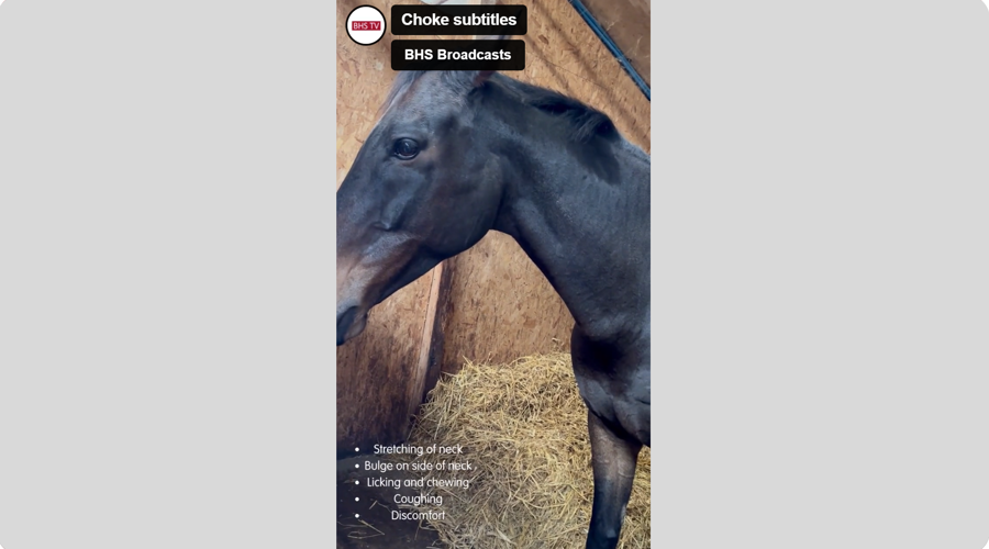
Diamond is a 12-year-old ex-racehorse with no previous history of health conditions and regularly sees an equine dental technician for routine work. Diamond’s symptoms were first spotted when coming into his stable in the evenings and after about half an hour of eating his hay he would go to the back of his stable and shake his head. Most would think he had gone to have a rest, but his owner quickly recognised this as an odd behaviour for him and observed more closely. Diamond would stretch his chin up as high as he could before then licking and chewing with his mouth (video below). This would happen for five or so minutes and then he would go back to his hay and start the whole process again.
 play-circle
Watch
This progressed to a large bulge forming at the base of the left side of his neck (photo below) accompanied by the signs listed above. The vet was called and did a thorough examination of Diamond’s mouth to check that he was able to chew properly, which all appeared to be normal.

Image shows an identifiable bulge on left side of neck.
The decision was made to sedate Diamond and pass a nasogastric tube into his oesophagus in the hope of dislodging any potential blockage that there may be. There didn’t appear to be any major blockage and no material travelled back up the tube. The instruction was to monitor Diamond carefully once the sedation had worn off to spot any signs of the bulge returning – which it did as soon as Diamond was offered his hay for the night.
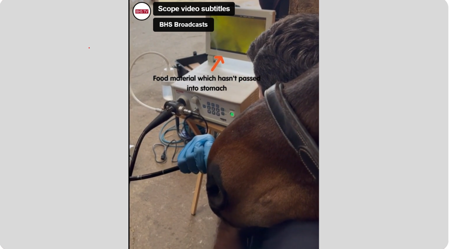
The next step was to use an endoscope to locate debris which may be stuck in the oesophagus or recognise any potential damage which had been caused. Sometimes damage to the oesophagus can form a ‘pocket’ which then allows food to become stuck, forming a lump. As you can see from Diamond’s endoscope footage below, there were no obvious signs of damage but a lot of food material that hadn’t travelled down the oesophagus and into the stomach. The vet also commented on a lack of motility, meaning that food material wasn’t getting pushed down by the muscles contracting.
 play-circle
Watch
Scope shows food matter in the oesophagus hadn’t passed to the stomach
After some research, Diamond was diagnosed with ‘distension of the distal oesophagus’ which means the lower section of his oesophagus has become stretched and isn’t pushing the food into the stomach, therefore causing a back flow. This condition is carefully managed and unfortunately cannot be cured.
Management practices:
“Diamond didn’t show signs of a ‘typical’ choke, but I knew something wasn’t right. On veterinary advice, I now offer all feed to Diamond from a height. Rather than give access to hay from the ground, I use a haynet and offer his hard feed from a bucket in his stable secured at chest height. This is to allow gravity to help encourage the material downwards. The haynet also slows down the intake of hay, preventing Diamond from swallowing too much at one time.
I’ve also found switching Diamond's feed to a mash extremely helpful as it helps to lubricate the digestive tract when eating his forage. I’ve also made sure that during winter when he spends more time in his stable eating hay, to split his feeds over three meals per day to help keep things moving. Due to his condition when he swallows the bulge still appears but then quickly goes again.
Over the summertime I was apprehensive about allowing Diamond to live out 24/7 as I was worried that with having his head down for most of the time, he may face an increase in choke episodes. However, after gradually introducing him to more time out in the field and making sure that the grass isn’t too long, he has coped perfectly with no episodes.
With a condition like this it’s important to recognise the subtle signs of abnormal behaviour and workout what works best for your horse. A lot of the practices I’ve put in place have come from trial and error to what suits Diamond best, and I have created a tailored plan with my vet regarding what to do if a choke episode occurs and when I need to call them.
Diamond is now back in full work and gaining condition while being able to be a horse with his friends in the field – which is all I could have hoped for.”
References
chevron-down
chevron-up
- Bell Equine Veterinary Clinic. (2023) Choke.
- Carson, D. & Ricketts, S. (2023) Choke in horses. VCA Animal Hospitals.
- Ridings Equine Vets. (2018) What to do if your horse gets choke.
- Haywood, L. (2022) Understanding choke in horses.
- Sutton, D. (2015) Diagnosing disorders of the equine oesophagus. 27(6). Pp. 291-294.
Get in touch – we’re here to help
The Horse Care and Welfare Team are here to help and can offer you further advice with any questions you may have. Contact us on 02476 840517* or email welfare@bhs.org.uk – You can also get in touch with us via our social media channels.
Opening times are 8:35 am - 5 pm from Monday – Thursday and 8:35 am - 3 pm on Friday.
*Calls may be recorded for monitoring purposes.


